
Ring 2: Technical Imaging and Scientific Research
Sequentially Shifted Excitation Raman Microscopy (SSE-RM) for Non-invasive, in situ Identification of Lake Pigments
Presentation by Kristen E. Watts, Ph.D., NRC Postdoctoral Fellow

Dr. Kristen E. Watts is a trained analytical chemist specializing in mass spectrometry and spectroscopy of organic materials. On January 13, 2021, as part of the Washington Conservation Guild’s 2022 3-Ring Circus, Watts presented the innovative research she conducted during her National Research Council (NRC) Postdoctoral Fellowship at the National Institute of Standards and Technology (NIST) from May 2021 through January 2022. The goal of her research project was to develop a non-invasive type of Raman spectroscopy that identifies organic red lake pigments using data processing focused methods which can be carried out in situ with relatively portable instrumentation. Due to the virtual nature of the conference, Dr. Watts was able to present from her office at NIST, while concurrently running an experiment.
She began her presentation with a brief history of the identification of red lake pigments, emphasizing that—while they are ubiquitous in cultural heritage—they are notoriously hard to identify in a non-invasive manner. She cites research published in 2006 by Whitney et al., in which surface enhanced Raman spectroscopy (SERS) was applied to identify red lakes.1 Prior to this study, Raman had not been employed to great effect for organic lakes because they are not efficient at scattering Raman light, and the data is also accompanied by large background fluorescence. Another type of analysis that is used is Fourier-transform Raman spectroscopy (FT-Raman). Watts explained the drawbacks to using SERS and FT-Raman, including that SERS is invasive because a nano-patterned silver substrate needs to be applied to the sample, and the equipment used for FT-Raman is complex and bulky—making portability and in situ analysis unfeasible.
Dr. Watts summarized key principles of light theory, explaining that changing the excitation wavelength will change the wavelength of subsequently Raman scattered light, but it will not change the wavelength of the background fluorescence (emitted light); it will only affect the intensity (amplitude) of the background fluorescence, not its position in spectral space. She also noted that artifacts of the experiment, such as fixed patterned detector noise and stray light, remain completely independent of excitation wavelength. In other words, stepping excitation—or gradual changes in the excitation wavelength—will only show changes in the Raman signal.
There are a number of techniques predicated on the premise of analyzing stepping excitation with data processing focused methods. Dr. Watts based her data processing on Sequentially Shifted Excitation Raman Spectroscopy (SSE-RS). In her experiments, Watts incrementally stepped the temperature of a diode laser to produce small changes in the excitation wavelength, thus inducing shifts in the obtained Raman spectrum. The datasets are processed using an algorithm that extracts the Raman signal from the fluorescent background. Due to time limitations, Watts was unable to go into specifics regarding how to code and apply the algorithms; for further information, she directed attendees to an article by Cooper et al.2 Her research credits the established precedence for using SSE-RS in cultural heritage3,4,5,6 but states that her work is distinct from published applications for the identification of organic pigments because:
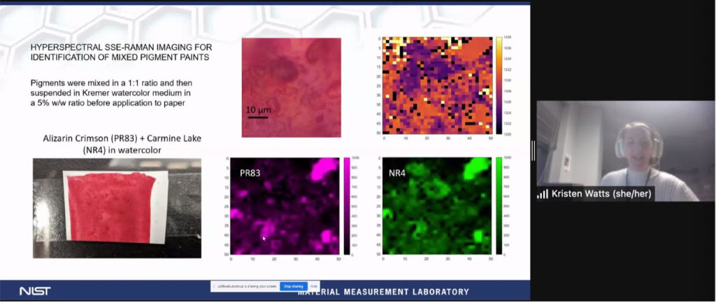
- She incorporates this technique into a microscope to engage in Raman hyperspectral imaging on a very small scale, yielding a spatial resolution for imaging objects in situ, as well as, discerning where pigment particles are relative to each other in the sample.
- Her research specifically monitors carmine lake and lac dye.
- The processing algorithm she coded is slightly modified from what is reported in the literature.
Dr. Watts walked the audience through her impressive microscope set-up and experiments using the equipment on pure pigment samples, paint-outs of single colors on paper, as well as mixed colors on paper. Successful tests indicate that Sequentially Shifted Excitation Raman Spectroscopy can be readily and successfully incorporated into a benchtop microscope for identification of natural red pigments for both point spectroscopy and hyperspectral imaging. Furthermore, SSE-Raman Microscopy can distinguish between different red lake pigment particles incorporated into the same paint on both a macroscopic and microscopic scale. Watts’ future direction involves translating this technique into a smaller portable system that could be used in situ using a mini-CCD and fiber optic probe technology.
References cited by Dr. Watts during her presentation:
(1) Whitney, A. V., Van Duyne, R. P., & Casadio, F. (2006). An innovative surface-enhanced Raman spectroscopy (SERS) method for the identification of six historical red lakes and dyestuffs. Journal of Raman Spectroscopy, 37(10), 993-1002. https://doi.org/10.1002/jrs.1576
(2) Cooper, J. B., Abdelkader, M., & Wise, K. L. (2013). Sequentially Shifted Excitation Raman Spectroscopy: Novel Algorithm and Instrumentation for Fluorescence-Free Raman Spectroscopy in Spectral Space. Applied Spectroscopy, 67(8), 973–984. https://doi.org/10.1366/12-06852
Notable applications of portable SSE-RS for point ID of organic pigments:
(3) Conti, C., Botteon, A., Bertasa, M., Colombo, C., Realini, M., & Sali, D. (2016). Portable Sequentially Shifted Excitation Raman spectroscopy as an innovative tool for in situ chemical interrogation of painted surfaces. Analyst, 141(15), 4599–4607. https://doi.org/10.1039/C6AN00753H
(4) Vagnini M, Gabrieli F, Daveri A, Sali D. Handheld new technology Raman and portable FT-IR spectrometers as complementary tools for the in-situ identification of organic materials in modern art. Spectrochimica Acta Part A: Molecular and Biomolecular Spectroscopy. 2017;176:174-182. https://doi.org/10.1016/j.saa.2017.01.006
(5) Pozzi, F, Basso, E, Rizzo, A, Cesaratto, A, Tague, TJ. Evaluation and optimization of the potential of a handheld Raman spectrometer: in situ, noninvasive materials characterization in artworks. J Raman Spectrosc. 2019; 50: 861– 872. https://doi.org/10.1002/jrs.5585
(6) Pause R, van der Werf ID, van den Berg KJ. Identification of Pre-1950 Synthetic Organic Pigments in Artists’ Paints. A Non-Invasive Approach Using Handheld Raman Spectroscopy. Heritage. 2021; 4(3):1348-1365. https://doi.org/10.3390/heritage4030073
WCG summary author: Sarah Mastrangelo, Contract Conservator for Paintings, Hirshhorn Museum and Sculpture Garden
Meeting attendance: 62 participants
