
Ring 2: Technical Imaging and Scientific Research
Application of Reflectance FTIR Spectroscopy for Insights into Photographs
Presenter: Dr. Joan Walker, Conservation Scientist at the National Gallery of Art (Washington, DC)
When collecting and analyzing scientific data of a complex surface, it’s imperative that the properties of the analyzed surface and the attributes of the instrumental technique are taken into careful consideration. Dr. Joan Walker really hit this point home in her talk for the second session of this year’s “3-Ring Circus,” during which she presented her findings from a research project comparing spectra collected of various photographic surfaces using attenuated total reflectance–Fourier transform infrared spectroscopy (ATR-FTIR) and portable external reflectance Fourier transform infrared spectroscopy (ER-FTIR). These two instrumental techniques are especially appropriate for collecting data on heritage materials, as they do not require sampling, a procedure that typically is not appropriate for photographs. Although ATR-FTIR requires contact with the surface (potentially leaving behind a small indentation on a deformable surface), the resulting spectra are generally straightforward to analyze: the technique is very surface-specific, penetrating approximately 1 μm into the sample. ER-FTIR, on the other hand, does not require contact, and the resulting spectra may contain specular (i.e., surface) and diffuse (i.e., volume) components, complicating interpretation.
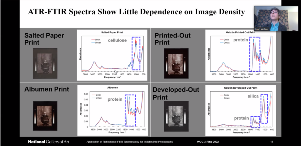
To establish how the spectra collected with these two FTIR techniques may vary, Dr. Walker analyzed a host of photographs from the study collections at the National Gallery of Art (NGA). The analysis of single layer photographs is quite straightforward: spectra of salted paper prints only show cellulose; spectra of cyanotypes show cellulose with and without contributions from the chemical signatures of Prussian blue, depending on the image density. However, the spectra collected of multi-layer photographs increase in complexity when probed with ER-FTIR: shiny subsurface layers (e.g., baryta in a gelatin print) do contribute to the obtained spectrum, whereas matte subsurface layers do not (e.g., paper support in an albumen print). The last portion of Dr. Walker’s talk highlighted how coatings on photographs can affect the resultant spectra. For example, characteristic bands of beeswax and cellulose can be identified in a spectrum collected with ATR-FTIR of a beeswax-coated salted paper print, but the cellulose signal is lost when ER-FTIR is employed.
Dr. Walker’s talk highlighted the immense degree of care that must be taken during the analysis of spectroscopic data. All aspects of a material’s chemical and physical properties must be considered, along with an understanding of how a probing light source will interact with different layers of a multi-layer material. Dr. Walker’s talk also reinforced the value of these types of comparative studies using study collections. Without the level of understanding gathered from studies like Dr. Walker’s, it’s far too easy to misinterpret spectroscopic data collected from complex or multilayer materials.
Meeting summary by: Dr. Teresa Duncan
Attendance: 63 participants
